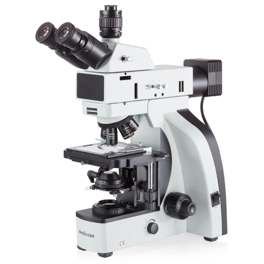
1. Overview
The AmScope T800 Series Infinity-Corrected Metallurgical Trinocular Compound Microscope is a professional-grade microscope designed for high-resolution inspection of metals, alloys, coatings, semiconductors, and other opaque materials. It features an infinity-corrected optical system, super-widefield eyepieces, a trinocular photo port for digital imaging, and both reflected (episcopic) and optional transmitted illumination. The T800 series supports multiple objective configurations and offers stable mechanical performance for routine laboratory work, research, and industrial quality control.
Capabilities
- The combination of high-quality optics, infinity-correction, and advanced illumination techniques ensures that the T800 Series delivers sharp, high-contrast images suitable for detailed analysis.
- Supports both transmitted (bottom light) and reflected (top light) illumination, making it suitable for both metallurgical and biological samples
- Polarizafion filter available
Features


2. Principle
- Infinity-Corrected Optical System: The microscope uses an infinity optical design, sending parallel light beams through the objective so the tube lens forms the final image. This system minimizes aberration and allows easy insertion of filters or polarizers without degrading image quality.
- Reflected (Episcopic) Illumination: Light from the epi-illuminator is directed onto the sample surface and reflected back into the objective, enabling detailed imaging of opaque specimens such as metals, circuit boards, and coatings.
- Transmitted Illumination (on supported models): Thin or semi-transparent samples can be viewed using a transmitted LED light source. Condensers and adjustable illumination help enhance contrast and resolution.
- Trinocular Phototube: The trinocular head includes a dedicated port for cameras, enabling image capture, real-time viewing, and digital measurement using imaging software.
3. Data Interpretation
- Magnification & Field of View: Choose different objectives to balance detail and viewing area. Low magnification provides broader context, while high magnification reveals finer microstructural features.
- Contrast Adjustments: Reflected brightfield highlights surface texture, scratches, grain boundaries, inclusions, and coating defects. Adjusting light intensity, angle, or filters enhances contrast and reveals additional structural information.
- Surface vs. Cross-Section Analysis: Reflected illumination gives information about surface morphology. Transmitted light supports examination of thin sections or mounted specimens for subsurface or internal features.
- Digital Imaging & Measurement: Images captured through the trinocular port can be used to measure grain size, crack width, coating uniformity, pore distribution, or defect density. Proper calibration ensures consistent, quantitative results.
4. Example Applications
- Metallographic analysis of grain structure, phase distribution, inclusions, and heat-treatment effects
- Inspection of solder joints, PCB surfaces, bonding pads, and micro-electronic features
- Evaluation of coatings, plating layers, corrosion, and wear patterns
- Research on alloys, ceramics, composites, and surface-engineered materials
- Failure analysis of fractures, fatigue surfaces, and micro-defects
- Educational training in materials science and metallography techniques